


Thoracic fibroids are rare in clinical practice. Previously, it was mainly diagnosed in the elderly, but now it is usually detected in patients under 35 years of age. Most often, the pathology develops in women than in men. This degenerative-dystrophy is difficult to diagnose, as severe symptoms appear only in a later stage.
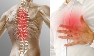
In addition, symptoms of this disorder can be easily confused with signs of impaired heart and lung function. The disease cannot be left untreated as it can cause scoliosis, development of persistent pain syndrome and other complications that can negatively affect a person's quality of life.
What is thoracic fibroids?
In the international classification this pathological condition has the code ICD-10 - M42. Thoracic tumors are much less common than the cervix or sacrum. This is no coincidence. Due to the presence of a rigid skeleton in this part of the body, this part of the spine is physiologically less mobile.
The thoracic region contains more vertebrae than the neck and lumbar, but in this part of the spine, the discs are thinner. These anatomical features contribute to reduced mobility of this part of the spine, resulting in less trauma.
However, when exposed to some adverse factors, osteonecrosis disease can develop. At first, there are signs of damage in a disc, but in the future, other factors may be involved in the pathological process. As the disease progresses, the bone elements as well as the ligaments and muscles that support the spine are damaged.
The breast degeneration is slower. Often years pass before the fibrous rings of the disc are so damaged that convex and herniated nipples appear.
Serious clinical manifestations have occurred after the height of the interlocking disc and roots decreased significantly. This may not only lead to low back pain, ie short-term pain in the chest area, but also lead to a violation of internal organs. It is much more difficult to treat the prolonged pinched nerve roots in this area.
Development reason
In most cases, spinal problems do not come suddenly. A disease such as osteonecrosis is no exception in this regard. This pathology, affecting the disc, is the result of long-term degenerative-dystrophy. In most cases, it is not possible to determine exactly what triggered the development of the disorder. Factors that can cause the appearance of osteonecrosis of the thoracic spine include:
- congenital or acquired malformation of the spine;
- overweight;
- overload of the spine during pregnancy;
- infectious diseases;
- hypothermia; metabolic disorder
- ;
- hormonal disorders;
- chronic stress;
- bad habit;
- connective tissue disease;
- dysplasia;
- postural disturbances;
- unhealthy diet;
- Injury.
Exercise negatively affects the condition of the spine. People with sedentary lifestyles are more likely to experience chest necrosis. In addition, age-related changes and a slowdown in metabolism, observed in patients over 55 years of age, contribute to the occurrence of these disorders.
Genetic predisposition may be a factor that can provoke the development of pathology. The genes that facilitate the occurrence of necrosis of the breast have not been determined, but people with a family history of the disease are more often diagnosed.
Symptoms and Signs
The clinic of this pathological condition depends on the stage of neglect of the process, the degree of damage to the disc that occurs, and the age of the patient. In the early stages of development, there are no specific signs, but general symptoms can occur cyclically. Usually, in the early stages of development, the disease manifests itself only at the onset of cold weather or after strenuous exercise. Early manifestations of necrotic growth of the thoracic region include:
- fast corrosion resistance;
- back pain and pressure;
- muscle spasms;
- cold limbs.
As the disease progressed, the patient's condition deteriorated. Chest pain appears. They are especially common in the background in a prolonged position or with sudden movements. In addition, severe pain syndrome can appear when lifting weights. Body rotation can increase pain. The presence of bone necrosis is also manifested by the appearance of dull pain in the shoulder area.
Usually, thoracic bone necrosis is accompanied by the appearance of an abnormal flexion. In severe cases, the patient may develop a lump. In addition, this disease can cause the appearance of pain when breathing deeply and exhaling.
When the nerve roots are pinched, there is usually a numbness in the limbs and skin on the body. Due to a violation of the endocrine and blood circulation, a sensation of goosebumps appears on the skin. Feet and hands are always cold. There may be a sensitivity to the limbs. In advanced cases, this disease can lead to the appearance of symptoms of damage to other organs due to a violation of their internal organs. In the final stage of the process, it is possible:

- intercostal nerve pain;
- stool disorder;
- flatulence;
- heartburn and nausea;
- itching and burning in the feet;
- violation of the reproductive system;
- asthma attack.
As the disease progresses, a person's ability to work decreases. Reduced physical activity. In the future, this disorder may set the stage for the development of serious complications. The risk of pathological fractures increases. The curvature of the spine leads to compression of the organs located in the chest.
With an unfavorable development, the disease continues to invade the myocardium and reduce lung volume. Often, such serious complications are accompanied by extensive bone necrosis, in which several intervertebral discs are affected at the same time.
Degree of thoracic necrosis
The existingclassification breaks down the development of this pathological condition into 4 degrees. Each of them is characterized by the presence of certain changes in the structure of the disc, vertebrae and other factors that form this part of the spine.
The first level
At the first degree of pathology, there is no obvious clinical manifestation, but specific changes in the structure of the disc can be detected with a comprehensive diagnosis. The fiber ring, which receives less moisture and nutrients, loses its elasticity. Small cracks often form in tissues in which the pulp is forced outwards. The disc can be moved into the spinal canal. Overhang is formed. There was no sign of ring rupture.
The second level
When the disease transitions to degree two, the first clinical manifestations are observed. Periodically, the patient suffers from pain and other neurological signs. When conducting specific diagnostics, signs of a decrease in the elasticity of the lumbar fibrous formation can be detected. The cartilage becomes thinner, leading to an increased risk of a hernia. There is a decrease in the height of the disc, as a result of which the structures of the spine have abnormal mobility.
The third degree
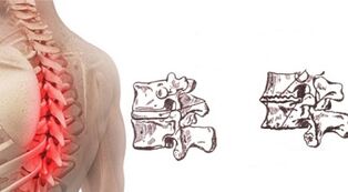
At the third stage, changes in the structure of the disc become so pronounced that the first signs of the development of kyphosis or scoliosis appear. Usually, at this stage in the process, the damaged loops are broken. This phenomenon is accompanied by the escape of the pulp nucleus outside the disc. Developing hernias, depending on the direction of the bulge, can compress nerve roots or spinal cord. Severe pain and nervous disturbances occur. Increased spinal mobility will facilitate injuries and fractures.
The fourth level
With the transition of the pathology to the fourth level of development, the structure of the discs is so disordered that they no longer perform an amortization function. The annular and nucleus fibrosus lose elasticity. These factors started to increase. Due to the violation of the decomposition function of the disc, the vertebrae are affected, subject to too much load.
At the edges of the vertebra adjacent to the damaged disc, osteogenous cells, that is, the growing bone, begin to grow rapidly. The surrounding ligaments are involved in the pathological process. They lose elasticity and no longer support the spine properly. In addition, at this stage of the development of the pathological process, the work of the muscle apparatus is interrupted.
Diagnosis
When signs of development of this disorder appear, the patient should be consulted with a neurologist and orthopedic surgeon. First, the doctor conducts an external examination and collects a history. Laboratory tests often indicated in the diagnosis of this disease include blood and urine tests. For information about the presence of defects in the structure of the spine, take an X-ray. This study reveals:
- reduces disc height;
- serrated edges of elements; herniation
- ;
- altered vertebral body;
- formed osteogenesis cells, v. v.
To clarify the defects in the structure of the disk, a disc is specified. This study allows you to identify the uneven core contours of the root canal, assess the extent of disc destruction and decrease tissue density. CT and MRI are usually done for better images. Considering that the clinical manifestations of thoracic necrosis are similar to those of myocardial ischemia, an electrocardiogram is often indicated to differentiate these conditions.
Treatment Options
This medical condition requires complex treatment. First of all, the patient is given a choice of drugs that help to eliminate symptoms and improve the nutrition of the disc. Drug therapy should be complemented by physical therapy and exercise. As a supplement, you can use some folk remedies. In addition, you should follow a specific diet.
Medicines
In the case of severe pain syndrome, the patient should adhere to a bed rest regime. This will reduce the intensity of the pain. To eliminate discomfort, pain relievers and NSAIDs are often prescribed. If the pain syndrome is too severe, a blockage method may be needed. Usually, glucocorticosteroids are prescribed to eliminate the pain in this disease.
Chondroprotectors are prescribed to improve the nutrient and water saturation of the disc. In some cases, antispasmodics and muscle relaxants are prescribed in short courses. These medications help reduce muscle spasms. If necessary, diuretics are prescribed to eliminate soft tissue edema. To improve the pinched nerve condition, patients need to supplement B vitamins.
Physiotherapy and massage
Exercise therapy and massage are the most important ingredients of bone necrosis treatment, but they can be used only after medication suppresses symptoms. Correctly selected exercises improve lung ventilation and strengthen the muscles that support the spine.
First, all the necessary exercises must be learned under the supervision of a therapist. In the future, the patient can do the exercises at home. People with this condition may be advised to take classes in the pool.
Massage helps to remove hypertonic muscles and improves soft tissue nutrition. In order for the procedures to be harmless, they must be performed by a specialist. In most cases, classical massage is done, which involves rubbing, smoothing, and pinching the problem area. Acupressure and segment massage can have many benefits. These techniques involve acting on pain points. They help improve blood circulation and lymphatic drainage. In most cases, it is sufficient for the patient to do the procedure 2-3 times a week.
Acupuncture
This method involves placing needles on areas of the patient's body. This method allows you to quickly get rid of muscle pain and spasm. Acupuncture should be performed by an expert in this regard. If a specialist does this, the procedure will be almost painless. Acupuncture is contraindicated for people suffering from cancer diseases and mental disorders. This method should not be used to treat bone necrosis when there are severe inflammatory processes.
Manual therapy
Manual therapy restores the correct anatomical position of the vertebra. In addition, this method also helps reduce the intensity of pain and muscle spasm. This effect helps to restore the ligamentous apparatus. Such procedures can delay the development of this pathological condition. The timing of the course of manual therapy is individually selected for the patient.
Post-isometric relaxation techniques

The isotopic relaxation procedure is a special technique that involves stretching all the muscles around the spine and then relaxing them.
Such exercises should be performed under the supervision of a specialist, who can assess the correctness of the movement and the severity of the muscle tension. This method allows you to quickly eliminate pain and restore normal muscle and ligament function.
Folk remedies
It is not possible to treat osteonecrosis only with folk remedies, as this method can aggravate the course of the disease. It is best to use different formulations based on herbs and other natural ingredients to support traditional therapies. You should get your doctor's recommendations about the possibility of using this or a folk method before you start using it.
Celery root
Properly cooked celery root is said to help saturate cartilage nutrients and water. To prepare this remedy, 1 bulb needs to be chopped and poured with 1 liter of boiling water. You need to emphasize ingredients for at least 8 hours. After this time, you need to filter the product and take 1 teaspoon. 3 times a day before meals.
Sunflower roots
To treat cervical spine tumors, people often use the sharp water of sunflower roots. To prepare this product, you will need about 1 cup of chopped plant material, pour 3 liters of water. The mixture should boil for 3-5 minutes. After that, the agent needs to be cooled and taken in tea form for several days. To improve the taste of the drink, honey can be added. It is better to store the remainder of the medicine in a thermos flask.
Home ointment
A simple homemade ointment can be used for osteonecrosis. To make this product, you need to melt about 150 g of lard in a water bath. Then 2 tbsp should be entered there. l. natural wax.
Preparations must be boiled for at least 20 minutes. Then 1 tbsp should be added to the heated mixture. l. fir oil. Product needs to boil for another 20 minutes. Finally, 2-3 minutes before taking the can out of the heat, 1 tablespoon is added to the mixture. l. ammonia. The finished composition must be distributed in jars. Store homemade ointments in the refrigerator.
Nutrition for breast necrosis disease
Patients with thoracic necrosis should have a balanced diet. Adequate protein-rich foods should be included in the diet. It is recommended to regularly eat foods containing large amounts of chondroitin, including aspic from fish, jelly meats, etc. v. Mandatory inclusion of fermented dairy products, vegetables and fruits in the diet. Dishes should be steamed or grilled. Avoid greasy and fried foods. Should be eaten in small but frequent portions. This will help avoid overeating.
More serious: what to do?
During the acute phase of the disease, there is a need for a minimum reduction in activity. If possible, avoid poses that increase pain syndrome. First aid for exacerbations of osteonecrosis includes the use of medications that reduce the severity of edema, inflammation, and pain. The patient is advised to rest in bed. A diet should be followed during this period. Only after eliminating symptoms can you begin exercise therapy and physical therapy.
Forecast
Currently this disease can only be cured in the early stages of development. With a late diagnosis, therapy aims to eliminate symptoms and improve spinal mobility. In some cases, surgical treatment is required. With an integrated approach to therapy, a person with this condition can lead a holistic lifestyle without suffering from pain and other neurological disorders.
Precautions
To prevent the development of this pathological condition, you should avoid sudden heavy lifting. You should always dress in accordance with the weather, avoid hypothermia. In addition, in order to prevent bone necrosis, one should resist hypotension and monitor posture. As part of preventing this pathology, you should eat properly and carefully monitor your weight.